Written by John C. Godbold, Jr., DVM and Jennifer F. Johnson VMD
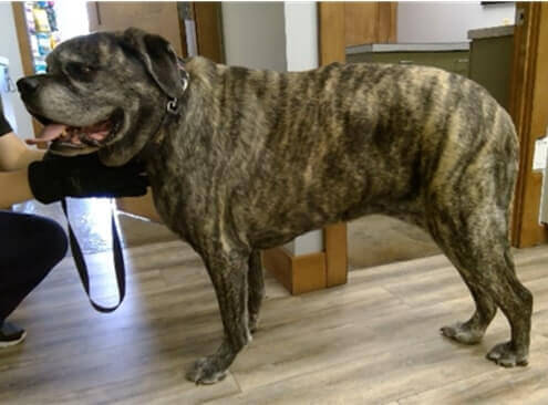
Sometimes, what veterinarians see with their eyes and feel with their hands, do not always tell the whole picture. A few thermal images can give us much more information. Meet Agadore Sparticus, a Bullmastiff with hindlimb lameness.
A Limping Patient
Agadore was an 8 year-old spayed female bullmastiff, who presented for right hindlimb lameness that had been observed for one month. On physical examination, she had a body condition of 4/5 and she slid her back legs along when she walked. Palpation of her back legs revealed pain over her right hip, and she had enlargement of her right metatarsals. The doctor also noted a dermal mass on her right flank that was draining purulent material, and another dermal mass on her dorsal thoracic area. Both masses were suspected to be sebaceous cysts.
Her thermal images showed thermal asymmetry with hyperthermia over the stifle, hock and tarsus in the right rear, which corresponded to the symptoms and physical findings. You can also appreciate significant hypothermia over the left hip. (Figure 1 and 2). It looks like everything is pointing towards arthritis of the right stifle and hock.
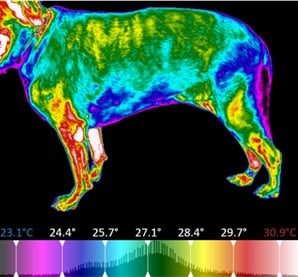
Figure 1 – left lateral
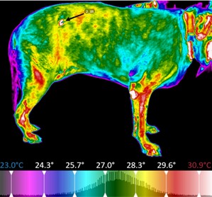
Figure 2 – Right Lateral
More Information is Gathered
In addition, the thermal images give us information about the dermal masses. The dorsal thoracic mass is visible as a discrete hypothermic area (Black arrow Figure 3), while the draining mass on the right lateral flank is hyperthermic (Black arrow, Figure 2). These are excellent representations which demonstrate the typical physiology of subcutaneous growths. Most benign, superficial growths have less vascularization and will be hypothermic, while infected, inflamed areas with increased circulation will show hyperthermia. Malignant growths are often hyperthermic as well. In this patient, the right lateral flank mass was definitively diagnosed as an infected sebaceous cyst, while the dorsal mass was a benign, sebaceous cyst.
Hypothermia is Significant

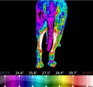
It is important to examine the entire patient when performing a physical exam, and the same rule applies to a thermal examination. In Agadore’s case, the final image offers more information, and speaks loudly (Figure 4). The posterior to anterior view of her hindlimbs revealed a large, focal area of hypothermia over the caudal aspect of the left thigh. The hypothermia is significant and decreased blood flow to the area offers a valuable clue. Nerve dysfunction, or neuropathy, will result in a loss of sympathetic inhibition, which causes peripheral vasoconstriction. When evaluating thermographs, marked hypothermia such as we see in this patient, should suggest we look for a neurologic cause. This area of the hindlimb is innervated by the caudal cutaneous femoral nerve, which originates in the L7-S1 area of the spinal cord. This image indicates the need for a more in-depth neurological examination and further diagnostic images of the lumbar and sacral areas.
Seeing the Entire Patient
Agadore demonstrates the great benefit of using thermal imaging in veterinary practice. While the history and physical examination pointed towards osteoarthritis of the right hindlimb, thermal images showed us a bigger picture. This is a great case where the pet owner could understand the need for further diagnostics involving the lumbosacral spine. Pinpointing all areas of disease will aid in treatment and pain relief, allowing for better patient outcomes and quality of life.
Thanks to Lori Keeler DVM, Willow Park Animal Clinic, Calgary AB