Osteoarthritis – Feline
Published by
Merissa Stockton
on
Patient
Domestic long hair, FeS, 13 year-old, 16 lb.
History
The patient had a history of reduced activity and not jumping on furniture anymore.
Physical Examination
A physical examination did not localize any sites of discomfort.
Physiological Screen with Digital Thermal Imaging
The patient was acclimated to room temperature, was not handled during acclimation, and remained calm during image capture. Digital thermal images were captured with a Digatherm IR camera.
Interpretation of the Thermal Images
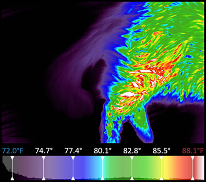
In the right hind limb increases in thermal gradients were noted within the soft tissues radiating out from the stifle joint, particularly over the sartorius and biceps femoralis muscles (Fig. 1).
In the left hind limb areas of hyperthermia were noted directly over the stifle joint and decreased in intensity as they radiate to surrounding soft tissues (Fig 2).
Additional Diagnostics
A radiographic study was recommended and the client readily complied.
Radiographs indicated early osteoarthritis of the stifles. (Figs. 3 & 4).
Treatment Recommendation
Weight loss, physical therapy, nutraceuticals, and laser therapy were prescribed.
Infrared Digital Thermal Imaging Provided
- Early detection of a disorder that was not detected during the physical exam.
- Client education and understanding, allowing immediate compliance to further diagnostics.
- Baseline information for monitoring this patient’s response to the treatment plan.