Pelvic Fracture – Feline
Published by
Merissa Stockton
on
History
This feline patient was presented for acute non-weight bearing in the rear quarters.
Physical Examination
The patient was extremely painful on physical examination and resisted any attempts to palpate the pelvis or rear legs.
Sedation for a physical exam was recommended but the owner declined.
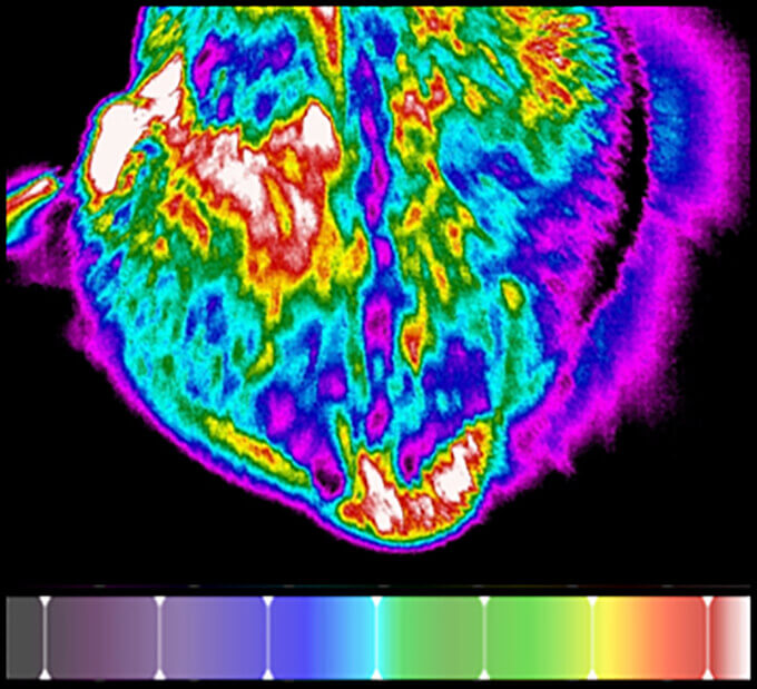
Regional Screen with Digital Thermal Imaging
Digital thermal images of the pelvis were captured with a Digatherm IR camera.
Interpretation of the Thermal Images
A dorsal image of the pelvis showed a region of asymmetrical hyperthermia over the left hip and the tail base (Fig. 1).
Additional Diagnostics
After seeing the thermal image, the owner agreed to sedation and radiographs. Further examination and radiographs indicated multiple pelvic fractures (Fig. 2).
Value of Digital Thermal Imaging
Client visualization of the digital thermal image contributed to compliance with the recommendation of sedation and radiographs.