Written by John C. Godbold, Jr., DVM and Jennifer F. Johnson VMD, CVPP
Thermal Imaging – helping veterinarians
Veterinary thermal imaging has quickly grown to be an indispensable tool for veterinary practice. With exceptional sensitivity, medically calibrated thermal imaging cameras exhibit such detail that they easily help clinicians see the unseen problems in their patients. Romeo is such a case.
Romeo was a nine-year old male neutered pit bull terrier dog, who presented in pain. He had a right cranial cruciate ligament (CCL) tear a year ago, which was surgically stabilized. Unfortunately, his left CCL suffered a complete tear 3 months ago, which had not had surgery.
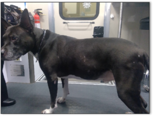
Physical examination revealed that Romeo was overweight (6/9), limping on his left hind, with cranial drawer present in the left stifle and pain localized to the left stifle. The right stifle exhibited decreased range-of-motion, medial buttressing of the stifle but no cranial drawer. When he rose from recumbency, he was slow to get up, using his forelimbs to pull himself up.
Surgical repair of the left CCL was not an option for these owners, and Romeo needed medical pain management. His veterinarian elected to incorporate photobiomodulation therapy (PBMT) into Romeo’s treatment plan. A series of thermal images were taken to perform a thorough whole-body evaluation.
When planning PBMT treatments, one needs to know where to treat. The thermal images helped to answer that question.
These lateral thermal images of Romeo demonstrate some expected findings when looking at a patient thermally. Higher temperatures are expected in any areas with shorter hair – the medial aspects of the forelegs, the axilla and the caudal abdominal and inguinal region. However, we see unexpected areas of increased temperature on the lateral aspect of both rear legs, over the shoulders, and on the posterior lateral aspects of both forelimbs below the elbows.
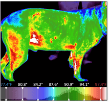
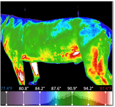
There is greater hyperthermia in the left rear, which is the stifle most recently injured. The hyperthermia in the right rear identifies inflammation likely due to off-loading the left rear. One can conclude the hyperthermia in the shoulders and antebrachia is due to postural compensation from offloading the hind end.
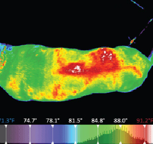
 This dorsal image of Romeo made the most impact on the PBMT plan, providing a complete picture of a key secondary area of concern. Noting the significant asymmetrical hyperthermia over the right thoracic and lumbar musculature, we identified additional areas of inflammation which contributed to the patient’s pain.
Thanks to Digatherm®, three simple images spoke for Romeo and helped to improve his care. His doctor better understood all the areas that needed to be treated. Without thermal images, we would not have recognized the compensatory changes in Romeo’s back and the need to include the back in the therapy.
Thanks to veterinary technician, Erin Shepherd in Lawrenceburg, IN for sharing this case.