Wondering how to make your lameness exams take less time and be more accurate in 2020?
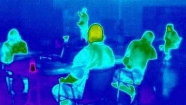
Adding thermal imaging to your exam toolbox will save you time by quickly pinpointing where the problems is and where your patient is compensating. Infrared thermal imaging (IRT) will give you a new way to get essential information during your lameness exams.
- A thorough medical history
- Visual assessment at rest
- IRT imaging of the entire patient
- Hands-on evaluation of the entire patient with special attention to areas of thermal asymmetry
- Through examination of all four feet including IRT of all four shoes and soles
- Visual evaluation while in motion
- Flexion tests
- Further diagnostic testing.
IRT is easy to include and images are quickly captured.
The images provide information about the physiological status of the patient in real time. Areas of hyperthermia are identified and show the extent of the anatomical area involved. Areas of hypothermia reveal vasoconstriction which usually indicate the need for a neurological evaluation.
Since horses are biomechanically balanced athletes, lameness disorders usually result in secondary compensatory issues. Including IRT in your lameness evaluations will allow early identification of all anatomical areas that will benefit from more detailed evaluation and diagnostic procedures.
IRT can provide images of the entire patient to the client so they understand and are compliant with your management recommendations. Thermal images help the client understand why you want to perform further diagnostics and why treatments are important. Once treatment has begun, thermal images monitor treatment progress and help establish when the patient is ready for a return to work.
Thermal Images Contribute to a Thoroughbred’s Lameness Exam
A 2-year old Thoroughbred was presented for evaluation of an acute lameness of the left fore. As part of the lameness exam, thermal images were captured.
A posterior to anterior image of the distal forelimbs pinpointed abnormalities. Areas of hyperthermia were noted medially along the entire length of the left metacarpus with a focal area at level 3. Focal hyperthermia was also noted proximal to the left medial sesamoid and over the left lateral sesamoid. An ultrasound study revealed core lesions within the suspensory ligament which correlated to the hyperthermia.
In this case thermal images helped focus diagnostics on an area of physiological change resulting in a quick diagnosis.
Schedule a demo today to see how digital thermal imaging will improve your lameness exams in 2020.


