Companion Animal

See the Unseen

Companion Animal Thermal Imaging

Companion animals are not able to show or tell us where their problems are. Digatherm®’s thermal imaging system solves that problem by letting us see the unseen and listen to what patients cannot tell us.

small-animal-thermography-infrared
small-animal-thermography-visual

Small Animal Thermography

Veterinarians utilize companion animal thermal imaging to see the unseen. First, a clinician must understand normal physiology in order to compare it to their patient’s current state.

Normal blood flow in cats and dogs results in symmetrical surface temperatures on both sides. Unhealthy patients can become non-symmetrical. At the same time, tissue changes can result in abnormal blood flow and non-symmetrical surface temperatures. Unexpected warmer or cooler areas, or asymmetry within the patient, distinguishes physiological change. Contralateral differences in temperature are also significant.

Increased temperatures (hyperthermia) are a direct result of increased blood flow. This may be due to inflammation, infection, or malignancy. Decreased temperatures (hypothermia) are a result of decreased blood flow. This is usually due to a neurological condition resulting in vasoconstriction. It is occasionally due to infarction or atrophy.

While thermal imaging cameras measure body surface temperatures, veterinary-specific software converts the temperatures into images. These thermal images use colors to represent the patient’s body surface temperatures. This provides a physiological map of the patient and in turn allows veterinarians to see the unseen.


spec

Medical-grade accuracy

spec

The pioneer in veterinary thermography

spec

Appropriate for every type of veterinary practice

spec

Comprehensive and individualized training


How do companion animal veterinarians use Digatherm®?

Three Ways

Leverage a thermal imaging camera for these three veterinary uses.

Evaluate Healthy Patients to Find the Unhealthy

Annual wellness evaluations are an important part of veterinary care. Veterinary patients are often stoic, and pet owners usually miss subtle signs of illness. Preventive care is proactive health. Digatherm® helps clinicians perform the most thorough assessment possible. Veterinarians use thermal images to collect baseline information during an examination.

Trained veterinary staff quickly take a series of images to evaluate the whole patient. Anything that appears abnormal is further investigated using palpation, anatomical imaging, or other diagnostic testing.

The sensitivity of thermal imaging provides a great addition to the physical examination. This guides the practitioner to look more closely and collect more specific clues.

Evaluate Unhealthy Patients

We know there is a problem. Let’s find it! Thermal images give a clearer and more concise way to a diagnosis. Many pets hide where they hurt. Thermal imaging helps find where and establishes a clearer, more concise diagnostic plan. The images pinpoint areas of concern and identify secondary compensation that would have been overlooked. This creates a more complete picture, especially when paired with anatomical diagnostics such as radiographs or ultrasound.

Monitor for Action and Reaction

The use of your Digatherm® does not stop after examination and diagnosis. Every problem requires a plan; Digatherm® is the first visual tool that shows us if the plan is working. Objectively assessing a patient’s response to treatment is difficult. It is hard to decide if something is working. Thermography measures progress and illustrates it to pet owners.

Communicate Better with Thermal Imaging

Digatherm® is a dream diagnostic tool in today’s technology-focused world. Pet owners understand and are comfortable using technology with their pets. They love the idea that the veterinarian can look closer and see the unseen. The bright, colorful images help pet owners visualize concerns and create a clear picture of any diagnostic plan. Pet owners want to educate themselves. Information builds trust and compliance.

Small-Animals-use-case-4

testimonial-quotation-icon“It has helped me be a better diagnostician by finding not only the areas of pain that are obvious,
but also the areas that are referred pain or secondary.”

– KERI GARCIA DVM, MRCVS

What can you see?

Below are several case examples of veterinary thermography for companion animals.

Case 1: Compensatory Inflammation

Software-Image-Digather-Resize-414x275-02romeo-leftromeo-rightromeo-pit-bull_visible


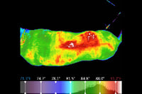
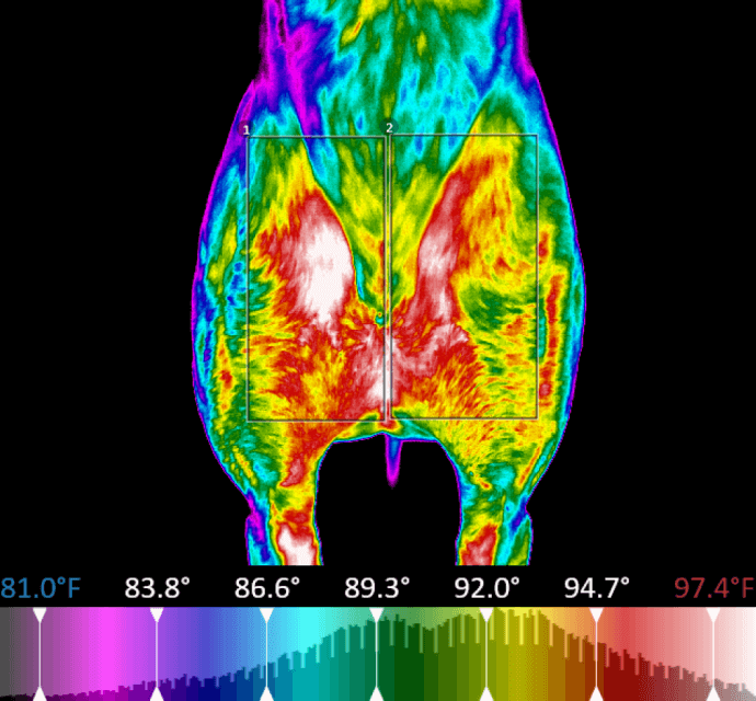
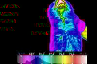
Presentation: Asymmetrical Hyperthermia Over the Dorsal Epaxial Muscles

Interpretation: This 9-year old Pitbull had bilateral cranial cruciate ligament tears. The area of asymmetrical hyperthermia over the dorsal expaxial muscles defines an area of compensatory inflammation in those muscles.

Credit: Erin Shepherd, RVT, On a Roll House Calls (Lawrenceburg, Indiana)

Case 2: Feline Hyperthyroidism

Software-Image-Digather-Resize-414x275-05

Presentation: Focal Hyperthermia Ventral Neck

Interpretation: This young adult domestic shorthair has a focal area of hyperthermia on the ventral aspect of the neck. A thyroid profile confirmed hyperthyroidism.

Credit: Kim Hunter, DVM, Vet to Pet Mobile Services (Smithers, British Columbia

Case 3: Possible Issues with Cervical Spine

Software-Image-Digather-Resize-414x275-08


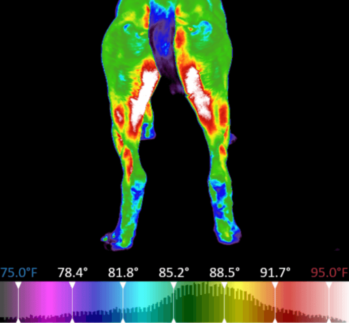
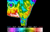
Presentation: Asymmetrical Hypothermic Dermatome Pattern

Interpretation: This ~3-5 year old American Bulldog/Pitbull Terrier has a focal area of hypothermia over the right carpus. This dermatome is innervated by the lateral branch of the superficial radial nerve which originates from the posterior cervical spinal segments. The image indicates the need for an in-depth neurological examination and further diagnostic imaging of the cervical spine.

Credit: Stephanie Schlachter, DVM, West-MEC (Glendale, Arizona)

Case 4: Benign Skin Mass

Software-Image-Digather-Resize-414x275-11


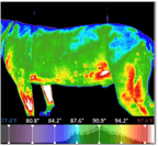
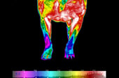
Presentation: Focal Hypothermia Lateral Thorax

Interpretation: This 6-year old Greyhound has a discrete focal area of hypothermia over the right lateral thorax that corresponds to a benign skin mass. Benign masses often have reduced vascularity and are hypothermic. Malignant masses often have increased vascularity and are hyperthermic.

Credit: Lauren Bueter, BS, RVT, Middlebury Animal Clinic (Middlebury, IN)

Case 5: Abnormal Weight Distribution

Software-Image-Digather-Resize-414x275-17


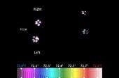
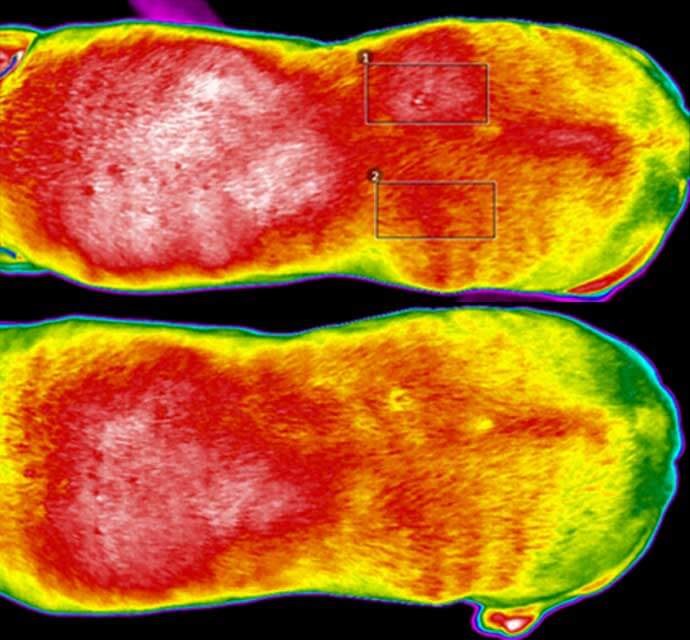
Presentation: Orthostatic Stance Analysis

Interpretation: This image of thermal paw-prints of a canine patient shows significantly higher thermal intensity of the fore paw prints with higher intensity on the left side giving a qualitative evaluation of weight bearing. The image suggests off loading of the rear legs and right side.

Credit: Stephanie Schlachter, DVM, West-MEC (Glendale, Arizona)

Case 6: Response to Therapy for Forelimb Lameness

Software-Image-Digather-Resize-414x275-20Software-Image-Digather-Resize-414x275-23


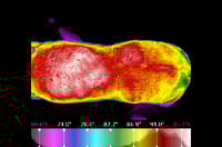
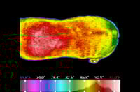
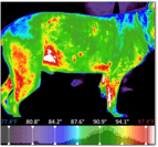
Presentation: Reduced Hyperthermia after Photobiomodulation Therapy

Interpretation: This first image of the back of a geriatric Boxer show hyperthermia over the dorsal thorax associated with compensation secondary to forelimb lameness. A similar image after three weeks of photobiomodulation therapy shows response to therapy with reduced hyperthermia and inflammation.

Credit: Mariah Richard, CVT, Barnesville Animal Clinic (Barnesville, Minnesota)

Digatherm® Thermal Imaging Systems

Learn more about Digatherm by ICI's veterinary tools and technology.

IR Pad 640

Our award-winning IR Pad 640 provides medical-grade resolution, accuracy, and sensitivity to the veterinary market. Veterinary specific software and calibration provide high-quality thermal images that are easily interpreted.

IR Pad 320

Our IR Pad 320 is easy to use in any veterinary practice. The handheld tablet allows vets to quickly visualize and document changes in a patient's physiological state in real-time.

https://infraredcameras.com/industries-served/veterinary/companion-animal/

Request a demonstration.

Discover the Digatherm by ICI difference and learn more about the most innovative and accurate thermal imagers available. Fill out the form below to request a demonstration of our state-of-the-art thermal camera.