Mixed Mammary Tumor – Canine
Published by
Merissa Stockton
on
Patient
English Setter, FeS, 5 year-old
History
The patient had previously been diagnosed with bilateral coxofemoral and stifle joint discomfort. Digital thermal imaging was initiated to establish a base line for monitoring this patient’s joint discomfort.
Regional Screen with Digital Thermal Imaging
The patient was acclimated to room temperature, was not handled during acclimation, and remained calm during image capture. Since the focus of the images was the hindquarters the patient’s bandanna was not removed.
Multiple digital thermal images were captured with a Digatherm IR camera.
Evaluation of the Right Lateral Thermal Image
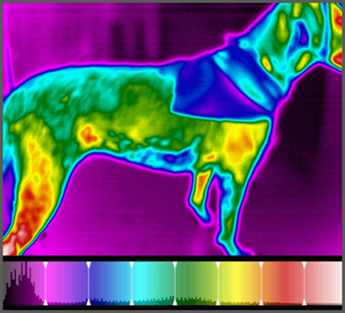
An area of decreased thermal gradient corresponds to the patient’s bandanna. An area of hyperthermia over the right stifle was expected since there was a previous diagnosis of early arthritic changes in the stifle.
An unexpected area of hyperthermia was noted along the ventral body wall anterior to the right flank. It corresponded to a palpable mass that had not previously been detected (Fig. 1).
Additional Diagnostics
The mass was surgically excised. Histopathology demonstrated the mass to be a mixed mammary tumor.
Value of Digital Thermal Imaging
Digital thermal imaging confirmed pathology in the right stifle and detected a previously undetected mixed mammary tumor.